Core Eye Services
Retinal Conditions and Treatments
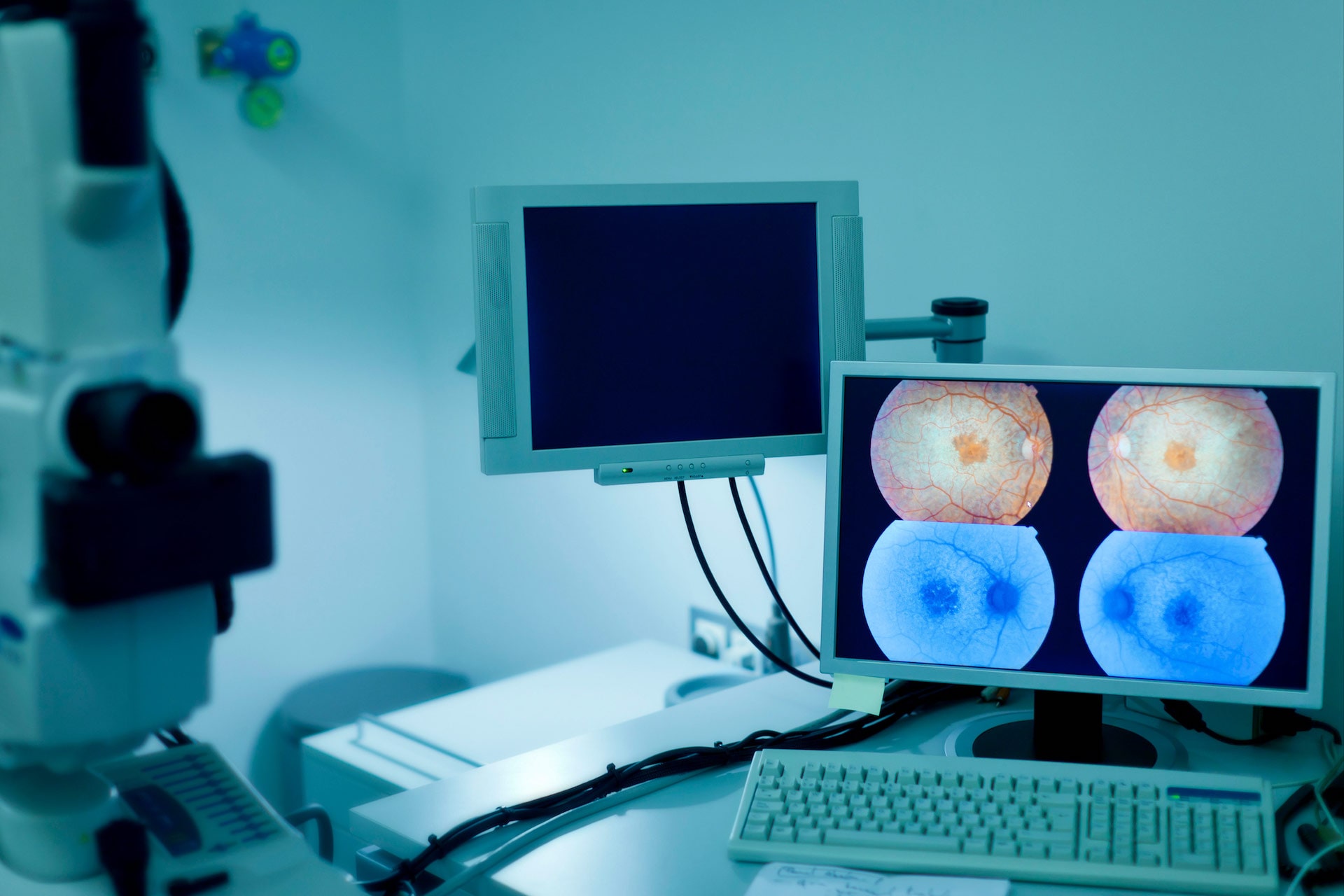
What is the Retina?
The retina is the layer at the very back of the eye whose function is to convert light entering the eye into electrical signals. These signals travel through the optic nerve and into the brain to create the images we see. It is like a translator in your eye – when light hits it, the retina converts it to a signal the brain understands.
The retina is made of two parts:
- Macula – located in the centre of the retina, it processes most of what the eye is looking directly
- Peripheral retina – it gives us our side (peripheral) vision and night vision
Top Three Retinal Problems
While there are several diseases that can affect the retina (or part of it), the three most common issues that we treat at Clarity Eye are:
- Diabetic retinopathy
- Macular degeneration
- Floaters and retinal tears
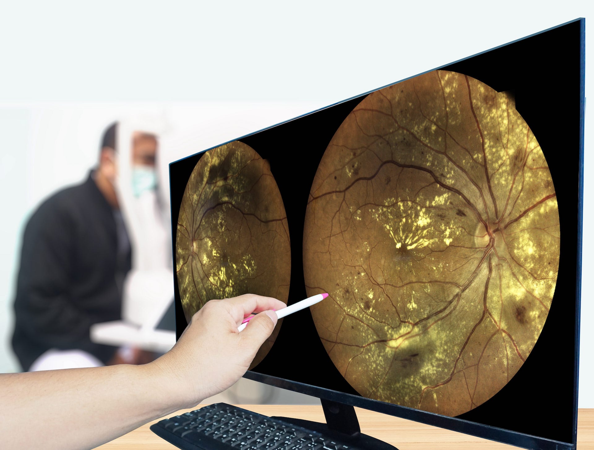
Diabetic Retinopathy
Diabetic retinopathy is a diabetes complication caused by blood vessel damage to the tissue at the back of the eye (retina). Initially, there may be no symptoms or only mild vision loss, but it can lead to eventual blindness. The condition can develop in anyone with type 1 or type 2 diabetes. The longer someone lives with diabetes and the less controlled the blood sugar levels, the more likely this eye complication will develop.
Diabetic Retinopathy Symptoms
As the condition develops, a person with diabetic retinopathy may encounter:
- Spots or dark strings floating in your vision (floaters)
- Blurred vision
- Fluctuating vision
- Dark or empty areas in your vision
- Vision loss
Macular Degeneration
Age-related macular degeneration (AMD), also known as macular degeneration, is a common eye disorder caused by deterioration of the macula - the small area in the centre of the retina in the back of the eye. Macular degeneration causes central vision loss - what you see directly in front of you.
Macular degeneration doesn’t cause total blindness because it doesn’t affect your peripheral vision.
There are two types of AMD:
- Dry macular degeneration - the most common type, affects 85-90% of individuals with the condition. It occurs when small yellow deposits, called drusen, develop under the macula.
- Wet macular degeneration – less common, but it occurs when abnormal blood vessels develop under the retina and macula
Risk Factors for Age-Related Macular Degeneration
There are several risk factors that can contribute to developing age-related macular degeneration, including:
- Age 65+
- Smoking
- High blood pressure or hypertension
- A diet high in saturated fat
- Race
Symptoms of Macular Degeneration
Macular degeneration is a progressive disease meaning the condition will worsen over time. Vision problems may go unnoticed in the early stages of the disease and changes may not be detected when both eyes vision loss occurs at the same time. Individuals may experience symptoms differently. Symptoms may include:
- Blurry or fuzzy vision
- Difficulty recognizing faces
- A reduction in central vision
- Straight lines appear wavy or distorted
- The need for brighter lighting
- Difficulty adapting to low lights
Wet macular degeneration symptoms resemble those of dry macular degeneration, such as visual distortions and reduced central vision but other symptoms experienced may include:
- A dark spot in the centre of your vision due to blood vessels bleeding or leaking fluid
- Hazy vision
- Rapidly worsening symptoms
Wet macular degeneration usually progresses more quickly.
Floaters and Retinal Tears
Eye floaters are small dark shapes that float across your vision resembling spots, specks, threads, or even little cobwebs. They are basically tiny shadows on your retina that travel across the eye when the eye itself moves and dart away when you try to focus on them directly. Floaters are caused by age-related changes that occur as the vitreous (a jelly-like substance inside the eye) becomes more liquid. Floaters come and go and often don’t require treatment. However, if there is a sudden increase in eye floaters or they don’t go away – especially if there are light flashes or a loss in peripheral vision – these can be symptoms of an emergency that requires immediate attention.
Risk Factors for Floaters
Almost everyone develops floaters as they age, but if you fall into the categories below, you may be at higher risk:
- Very nearsighted
- Have diabetes
- Have had cataract surgery
- Age 50+
Sometimes floaters have more serious causes, including:
- Eye infections
- Eye injuries
- Bleeding in the eye
- Uveitis (inflammation in the eye)
- Vitreous detachment (vitreous pulls away from the retina)
- Retinal tear (vitreous detachment tears a hole in the retina)
- Retinal detachment (retina gets pulled away from the back of the eye)
When to Get Immediate Help
- Many more eye floaters than usual
- A sudden onset of new floaters
- Flashes of light in the same eye as the floaters
- Darkness/shadow on any side(s) of your vision
Retinal tear or detachment is a sight-threatening condition that requires immediate attention. Go to your eye doctor or the emergency room immediately.
Treatments of Retinal Conditions
The two most common retinal treatments performed at Clarity Eye are:
- Intravitreal injections
- Retinal Argon laser

Intravitreal Injections
Just as it sounds, medicine is injected into the vitreous (the jelly-like fluid at the back of the eye) of the affected eye using a small needle. The medicine is used to treat a variety of retinal conditions including age-related macular degeneration (AMD), diabetic retinopathy and retinal vein occlusion. In some cases, an injection is used to insert a small gas bubble to aid repair of a retinal detachment. This method is most often used to get a higher level of medicine to the retina.
What to Expect
It takes about 2-3 minutes and while you may feel some pressure, the procedure is painless:
- Numbing drops are placed in the eye
- The eye is sterilized with iodine drops
- A small device is placed to keep the eyelid open during the procedure
- A small amount of medicine is injected into the eye with a small needle
- Antibiotic drops may be placed afterwards
Retinal Argon Laser
Retinal Argon laser (photocoagulation) is an in-office procedure used to treat numerous retinal conditions, including retinal tears, diabetic retinopathy, macular edema and retinal vein occlusion. It’s most frequently used to repair a retinal tear to prevent development of a retinal detachment, a potentially blinding condition. The surgeon directs a laser beam into the eye through the pupil which makes burns around the retinal tear, creating scarring that welds the retina to existing tissue. The procedure cannot typically restore vision already lost, but it can reduce your risk of experiencing future vision loss.
What to Expect
It takes about 5-10 minutes and while you may experience bright flashes of light or feel a slight dull ache as the laser is applied, the procedure is relatively painless:
- Drops are placed in the eye(s) to numb and dilate pupil(s)
- A special contact lens may be placed to precisely focus the laser
- Laser is used to photocoagulate the targeted areas
As eyes will remain dilated following the retinal laser procedure, you will need to arrange for someone to drive you home. Your vision may be blurry, and you may experience mild dull ache in the treated eye(s) for a few hours following the procedure. You may be advised to limit certain activities for a period of time following the procedure.