
Sudden, painless blurring of vision in one eye is a common reason patients get referred to our retina service. One of the leading causes behind it is retinal vein occlusion. It's a condition many people have never heard of until it happens to them. Yet it's one of the more common vascular causes of vision loss in adults over 50. Knowing the retinal vein occlusion symptoms early can help protect your long-term vision. It can also help prevent complications down the line.
What is Retinal vein occlusion?
Retinal vein occlusion (RVO) happens when a vein that drains blood from the retina gets blocked. Usually, this is caused by a blood clot. When blood can't drain properly, pressure builds up in the smaller vessels feeding into that vein.
These vessels then leak blood and fluid into the surrounding retinal tissue. This leads to swelling, especially in the macula. The macula is the central part of the retina responsible for detailed vision. It also reduces blood flow to parts of the retina that depend on that vein.
There are two main types. The difference matters for both symptoms and long-term outlook.
- Branch retinal vein occlusion (BRVO) – a blockage in one of the smaller branch veins. It feeds only a portion of the retina. Because only part of the retina is affected, vision loss is often less severe and more localized.
- Central retinal vein occlusion (CRVO) – a blockage in the main retinal vein itself. This affects blood drainage from the entire retina. It tends to cause more widespread swelling. In many cases, it causes more significant vision loss.
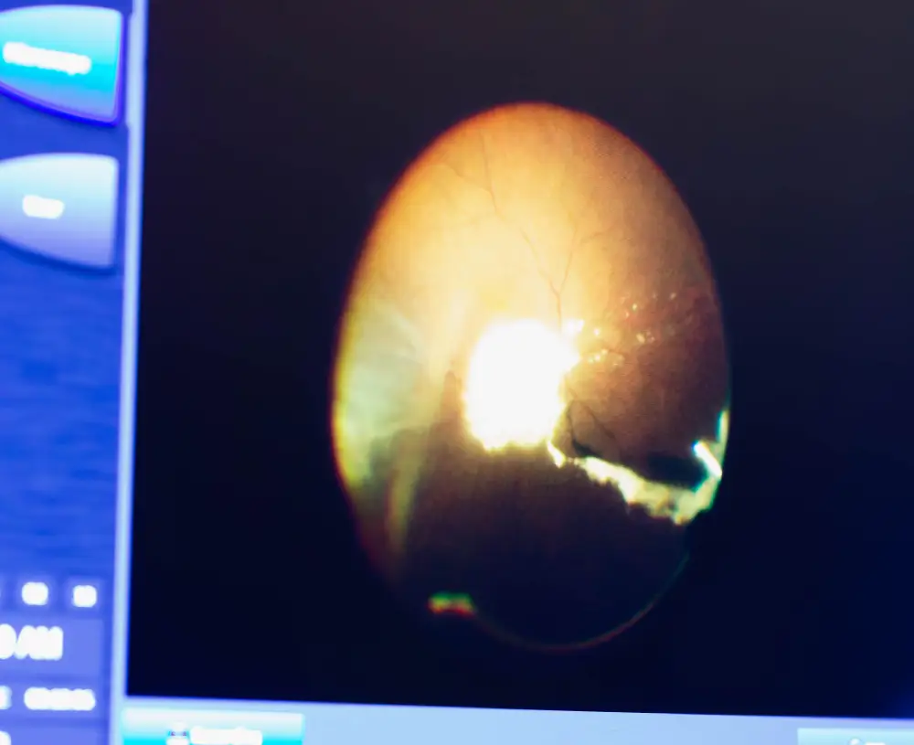
What are the symptoms of RVO?
The main symptom of retinal vein occlusion is sudden, painless blurring or dimming of vision. It almost always affects just one eye. Beyond that, patients often describe:
- A sense that part or all of their vision has become hazy, or "washed out"
- Distorted or patchy areas within their field of vision
- New floaters, especially if bleeding has happened inside the eye
- In more severe cases, a noticeable drop in vision. This happens over hours, not gradually.
RVO is almost always painless. If it's a branch occlusion, the change can be subtle at first too. Because of this, many people wait before booking an appointment. But waiting allows swelling to get worse. It raises the risk of complications. Any sudden, unexplained change in vision, even without pain, needs prompt evaluation.
What causes retinal vein occlusion?
RVO develops when a blood clot forms inside a retinal vein. This most commonly happens where the vein crosses over a hardened or thickened artery. This crossing point becomes a pinch site. It restricts normal blood flow. This sets the stage for a clot to form. Several health and lifestyle factors raise the likelihood of this happening:
- High blood pressure – one of the strongest and most common risk factors. Sustained pressure damages and stiffens blood vessel walls over time.
- Diabetes – contributes to blood vessel damage throughout the retina. This adds to the effects of a vein blockage.
- High cholesterol – contributes to the arterial stiffening that leads to vein compression.
- Glaucoma – elevated eye pressure has been linked to a higher risk of vein occlusion.
- Age – RVO is most commonly diagnosed in people over 50. This is because vascular changes build up over decades.
Less commonly, RVO can be linked to blood clotting disorders, inflammatory conditions, or other systemic vascular diseases. This is why a full medical history is often part of the diagnostic process.
Is retinal vein occlusion related to diabetes or high blood pressure?
Yes, both are among the most significant and well-documented risk factors for RVO. Patients being treated for diabetic retinopathy or managing glaucoma are already at higher risk. They benefit from regular retinal screening as part of their ongoing eye care, even without new symptoms. The National Eye Institute notes that managing these underlying vascular conditions matters a lot.
This includes blood pressure control, blood sugar management, and cholesterol treatment. Together, these reduce both the initial risk of RVO and the chance of it coming back or affecting the other eye.
This is also why RVO care rarely stops at the eye itself. Your ophthalmologist may recommend follow-up with your family doctor. This helps review blood pressure, blood sugar, and cardiovascular health. RVO can sometimes be an early signal of broader vascular issues that haven't been diagnosed yet.

How is RVO diagnosed?
Diagnosis starts with a full dilated eye exam. This lets your ophthalmologist directly examine the retina. They look for signs of bleeding, swelling, or vessel changes typical of RVO. From there, two imaging tools are usually used:
- OCT (optical coherence tomography) – a non-invasive scan that produces a detailed cross-section of the retina. This lets your doctor measure how much swelling there is, especially in the macula.
- Fluorescein angiography – a test using a special dye injected into the bloodstream. It highlights how blood is, or isn't, flowing through the retinal vessels. This helps identify blocked areas and abnormal vessel growth.
Together, these tools help figure out which type of RVO is present. They show how severe the swelling is. This helps decide which treatment approach fits best.
How is retinal vein occlusion treated?
Treatment for RVO focuses on two goals. First, reducing swelling to protect or restore vision. Second, preventing longer-term complications like abnormal blood vessel growth. Depending on severity, treatment typically involves:
- Intravitreal injections – medication injected directly into the eye. This reduces swelling and blocks the growth of abnormal blood vessels. This is currently the main treatment for macular swelling caused by RVO. It's explained in more detail on our retina services page. Injections are often needed on a regular schedule at first. They taper off as the eye responds.
- Laser photocoagulation – in some cases, laser treatment addresses areas of poor blood flow. It can reduce the risk of abnormal vessel growth, particularly in more advanced CRVO cases.
- Managing underlying conditions – working with your family physician to control blood pressure, blood sugar, and cholesterol. This lowers the risk of the occlusion getting worse or coming back in either eye.
RVO requires prompt, ongoing management to protect your vision. Book a retina consultation at the Clarity Eye clinic nearest you, especially if you've noticed any sudden, unexplained change in your vision.
Can vision recover after RVO?
Many patients see real visual improvement with treatment. This is especially true when treatment starts soon after diagnosis, before prolonged swelling causes lasting damage to the macula. That said, outcomes vary a lot based on:
- Whether the occlusion is branch or central (BRVO generally has a better visual outlook than CRVO)
- How much swelling has developed by the time treatment starts
- How consistently follow-up injections or monitoring appointments are attended
- Whether underlying vascular risk factors are being managed well
For many patients, RVO becomes a condition managed over months to years. It's rarely resolved after a single treatment. That's why building a steady relationship with a retina specialist matters just as much as the initial diagnosis.
Frequently asked questions
What is the difference between BRVO and CRVO?
BRVO affects a smaller branch vein and only part of the retina. This usually causes milder, more localized vision loss. CRVO affects the entire retina's main draining vein. It typically causes more widespread swelling and bigger vision changes.
Is retinal vein occlusion an emergency?
It's usually not as immediately sight-threatening as a retinal detachment. But any sudden, unexplained change in vision should be checked within a few days. This prevents swelling and complications from getting worse.
Can retinal vein occlusion be prevented?
There's no guaranteed way to prevent RVO. But managing blood pressure, blood sugar, and cholesterol lowers your risk a lot. Regular eye exams help too, especially if you have known cardiovascular risk factors.
Does RVO always require injections?
Not always. Mild cases with minimal swelling may just be monitored closely. But most cases involving central vision loss do benefit from injection therapy.
What's the outlook for someone diagnosed with RVO?
Outlook varies by type and severity. But many patients maintain or regain functional vision with consistent treatment. Actively managing the underlying health conditions that contributed to the occlusion also helps.
Can RVO happen in both eyes?
It's uncommon for RVO to happen in both eyes at once. But having RVO in one eye does raise the long-term risk of it developing in the other. This is especially true if underlying vascular risk factors aren't well controlled.