A detached retina is one of the few true emergencies in eye care. It's also one of the most treatable, but only if it's caught in time. Every year, thousands of Canadians notice sudden changes in their vision. Some of these changes turn out to be early signs of retinal detachment. How quickly someone acts often decides whether their vision fully recovers. Knowing the retinal detachment warning signs early can make a huge difference. It can mean the gap between a routine same-day procedure and permanent vision loss.
At Clarity Eye, our retinal specialists see patients across the Greater Toronto Area every week. Many of them weren't sure if their symptoms were serious enough to act on. Clarity Eye has one of the largest retina teams in the province and some of the most comprehensive retina coverage in Ontario. This guide walks through what retinal detachment is. It explains what it feels like, who's most at risk, and most importantly, when you need to be seen right away instead of waiting.
What is retinal detachment?
The retina is the light-sensitive layer at the back of your eye. Its job is to turn light into electrical signals. These signals travel through the optic nerve to your brain. Your brain then turns them into the images you see. Think of the retina like film in a camera. If it isn't sitting flat and properly connected, the picture doesn't form correctly.
Retinal detachment happens when this layer of tissue pulls away from the support tissue underneath it. That support tissue keeps the retina nourished and working. Once detached, the retina loses its blood supply. It stops working properly.
Depending on how much of the retina is involved, and how long it stays detached, this can range from a minor issue to a cause of permanent vision loss.
There are three types of retinal detachment. Knowing which type you may have affects both treatment and outlook.
- Rhegmatogenous detachment: the most common type. It's caused by a tear or hole in the retina. Fluid seeps underneath it and slowly lifts it away from the eye wall. This is usually linked to age-related changes in the gel inside the eye.
- Tractional detachment: happens when scar tissue on the retina's surface tightens. It physically pulls the retina away from the back of the eye. This is more common in people with advanced diabetic eye disease.
- Exudative detachment: caused by fluid building up under the retina, but without any actual tear or hole. It's often linked to inflammation, tumors, or other eye diseases.
What are the early warning signs of a detached retina?
Most people notice one or more warning signs before a full detachment happens. Catching these signs quickly is the biggest factor in saving your vision.
- A sudden increase in floaters. These are small specks, strings, rings, or cobweb-like shapes. They drift across your vision. You'll often notice them more against a plain background, like a white wall or blue sky.
- Flashes of light (photopsia). These are brief flickers or streaks of light. They're most noticeable in your peripheral, or side, vision. Low light also makes them more visible.
- A gray shadow or curtain. This is a dark area that seems to creep in from one side of your vision. It doesn't go away on its own.
- Blurred or distorted vision. This kind of blur doesn't clear up with blinking or resting your eyes.
These symptoms usually show up in one eye first. Unlike many eye conditions, they can develop and get worse within just a few hours. That's exactly why quick evaluation matters so much.
What does a detached retina feel like?
This is one of the most common questions people ask about this condition. The answer often surprises people. Retinal detachment is usually painless. There's no aching. There's no pressure. There's no obvious physical feeling that something is wrong inside the eye. The only clues are the visual symptoms listed above.
This is exactly why so many people delay getting care. Without pain, it's easy to assume the problem isn't urgent. But the absence of pain doesn't mean the situation isn't serious.
Is retinal detachment a medical emergency?
Yes, without question. The longer the retina stays detached, the more retinal cells get damaged from a lack of blood supply. This lowers the chance of recovering full vision. This is especially true if the detachment spreads to involve the macula. The macula is the small central area of the retina responsible for sharp, detailed vision.
According to the American Academy of Ophthalmology, outcomes are much better when treatment starts before the macula gets involved. This window is often within the first 24 to 72 hours after symptoms begin. Once the macula detaches, even successful surgery may not fully restore central vision.
If you notice sudden floaters, flashes, or a shadow in your vision, don't wait to see if it passes. Contact an eye doctor or go to your nearest emergency room right away. Retinal detachment can't wait for a routine appointment.
Clarity Eye’s retina team includes medical and surgical retina specialists and provides care across multiple GTA clinics. Learn more about the retinal service and Clarity Eye’s doctors.
For referring optometrists
Clarity Eye’s Retina Express service is open to referring optometrists to expedite and triage emergency retina cases. The dedicated line is listed as available Monday to Saturday, from 12:00 p.m. to 8:00 p.m., at 416-663-3937. It is intended for retina referrals and urgent retina-related inquiries.
How is retinal detachment treated?
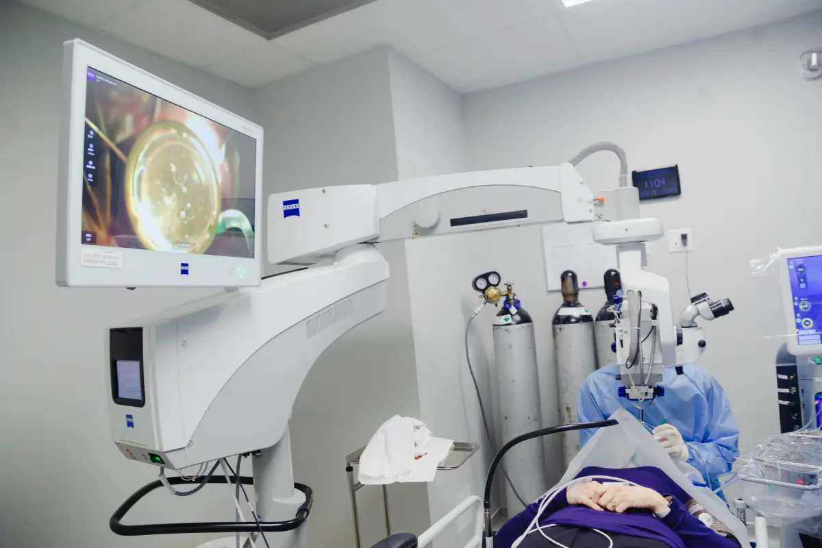
Treatment depends on a few things. It depends on the type, size, and location of the detachment. It also depends on how long it's been present. In many cases, doctors use a combination of approaches.
- Pneumatic retinopexy – a small gas bubble is injected into the eye. With careful head positioning, the bubble presses the retina back into place while it heals. This is often done as an in-office procedure for select cases.
- Scleral buckle – a flexible silicone band is placed around the outside of the eye. It gently indents the eye wall. This relieves the pulling force on the retina, letting it reattach.
- Vitrectomy – the gel inside the eye is surgically removed. It's replaced with a gas bubble, or less commonly, silicone oil. This gives the retina space and support to reattach properly.
Many patients with retinal tears are treated before a full detachment develops. They receive retinal laser photocoagulation. This seals the edges of the tear. It stops the tear from progressing further. Catching a tear at this stage often avoids the need for more invasive surgery.
Can a detached retina heal itself?
No, it can't. Unlike some minor eye irritations, retinal detachment won't resolve on its own. It needs prompt medical or surgical treatment. Delaying treatment, even by just a few days, can significantly lower your chances of a full visual recovery. In some cases, it can lead to permanent blindness in the affected eye.
Who is most at risk of retinal detachment?
Retinal detachment can happen to anyone. But certain factors raise your risk a lot.
- Age – most common in adults over 50. This is because the gel inside the eye naturally changes and separates from the retina over time.
- High myopia (severe nearsightedness) – longer eyes are more prone to retinal thinning and tears.
- Previous eye surgery – including cataract surgery. This can slightly raise detachment risk in the months or years after the procedure.
- Diabetes – especially advanced diabetic retinopathy. It can lead to tractional detachment.
- Eye trauma – a blow to the eye or head can cause a tear or detachment right away.
- Family history – having a personal or family history of retinal detachment raises your likelihood of experiencing one.
If you fall into one or more of these groups, routine dilated eye exams matter even more. They can catch early changes before you even notice any symptoms.
Frequently asked questions
How do you get a detached retina?
Most cases start with a retinal tear. This is often caused by age-related changes in the eye's gel pulling on the retina as it naturally separates. Trauma, high myopia, diabetes, and previous eye surgery also raise the risk.
Can a detached retina be fixed?
Yes, in most cases. Success rates for reattachment surgery are high, especially with early treatment. But how much vision you recover depends on how much of the retina, especially the macula, was affected before treatment.
What happens if retinal detachment goes untreated?
Untreated retinal detachment leads to progressive, and eventually permanent, vision loss or blindness in the affected eye. This happens as retinal tissue is starved of its blood supply.
How long does recovery take after retinal detachment surgery?
Most patients need several weeks for full visual recovery. But initial healing and improvement are often noticeable within the first few days to a couple of weeks.
Is retinal detachment surgery painful?
Most procedures use local anesthesia, so there's minimal discomfort during surgery itself. Some mild soreness, pressure, or scratchiness is normal for a few days afterwards.
Will my vision go back to normal after treatment?
It depends on how much of the retina, especially the macula, was involved before surgery. Many patients regain excellent vision. Others may notice some permanent changes if the macula was detached for a long time.